


订单编号
患者姓名
Company News Home > Company News
How to judge the dental colorimetric principle
How to judge the dental colorimetric principle
The current colorimetric principle in dentistry is based on the theory of the American painter Albert Henry Munsell in 1898, and Clark applied Munsell's theory to dentistry in 1930. According to this theory, color consists of three dimensions: hue, color, and lightness.
Hue is the basic color of teeth, color is the saturation degree of tone, and lightness represents brightness. The standard VITA color comparison board has four basic colors (A, B, C, D), and each tone has four shades.
In fact, tooth color is a combined result of the interaction of many factors. During the refraction and reflection of light waves, tooth enamel and dentin interact with light. In the enamel area, the short-wave light near white-blue light is dominant, while in dentin, the longer wavelength yellow-orange light is more obvious.
Dental color is a combined result of several factors, which must be carefully analyzed to understand the unique characteristics of each patient's teeth. The traditional method is not precise enough to achieve this.
To determine the tooth color, it is necessary to abandon the traditional color plate and adopt the personalized color analysis theory with higher tooth color analysis ability: Dr. Vanini's color theory. The tooth color that we usually see is the result of the physical features of dentin and enamel and their interaction with light.

Vanini's theory of tooth color includes a detailed analysis of each factor. Record each factor with a special color form. These factors are then reproduced using specific materials during the hierarchical filling stage.
Vanini's theory of tooth color includes a detailed analysis of each factor. Record each factor with a special color form. These factors are then reproduced using specific materials during the hierarchical filling stage.
To correctly determine the tooth color, the dentist must be able to see the deep depth of the tooth structure and identify five color dimensions and ribbons. To do this, we need a color temperature constant in 5500 kelvin light, there are several studies show that the light is the ideal evaluation color, then, using digital photography analysis color dimension, because digital photography technology can let us use the computer to teeth deeper inspection, more accurately determine different color dimensions. Insufficient image exposure and increased contrast will help you to better see the color dimension, and increase the shiny amber and blue colors at the cut end.
All tooth color information must be recorded in a simple way, for which we designed a special color chart for studying and determining the five color dimensions and the specific materials needed to obtain these effects. Color chart represents the repair scheme, strive to achieve the color chart scheme to achieve satisfactory repair effect. The details of the table also include two blue dental areas. On the left are the five color dimensions, and on the right are the initials of the composite resin material (i. e., enamel, dentin composite resin) needed to reproduce the color.
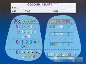
Figure: Color navigation table
At the back of the chart with strong white light, light, and characteristics of the classification, each dimension associated with age, each biotype prompt common dimensions of color and color saturation, to determine the first color dimension is basic color (BC), from the average of dentin color, should be by layered filling the same composite of the color plate in a third of the tooth color to determine. The basic chroma is recorded on the left side of the chart and the required dentin composite was recorded on the right. The shape and contour of the dentientine also need to be determined and reproduced.
Beauty plastic resin color comparison principle
The second dimension of enamel to be recorded is the lightness or brightness of the enamel, high lightness of the young biotype (UE 3), moderate lightness of the adult biotype (UE 2), and the old biotype (UE 1). This assessment can be done by taking black and white photographs.
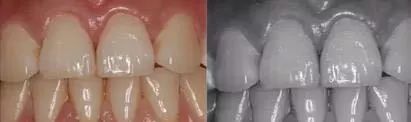
Doctor case sharing ↓↓↓
In order to determine the strong white light, milky white light and characteristics, compare the photos with behind the color chart, under-exposure and high contrast can help to analyze the image. Strong white light is mainly found in young biotypes, usually with type 1 (dots) and type 3 (snowflakes) seen. Adult and older biotypes are more common in type 2 (Xiaoyun) and type 4 (horizontal zone).
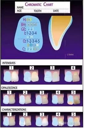
Young biotypes showed grey-blue tone of type 1 (mastoid) and type 2 (split mastoid); adult biotypes showed grey-blue tone of type 3 (comb tooth) and type 4 (window). Elderly biotype showed type 5 (stain t) amber tone.
Young biotypes are most commonly characterized by the mastoid process (type 1), which may be white or amber, forming a clear dividing line from the milky light; the cut margin (type 3) has white or amber lines. Common characteristics of older biotypes are white or amber one or more horizontal bands extending to the adjacent zone, where cut amber or brown stain-like features and enamel cracks (type 5) can be reproduced with brown cracks or white opaque cracks. Adult biotypes can emerge in all five traits.
Hot newsMore
- [07-07]全口义齿的美学修复特点
- [07-07]Restoration of
- [07-07]Choose from fou
- [07-07]Nursing care of


Copyright 2021 Shenzhen Deke dike Business Management Co., Ltd. all rights reserved
 粤ICP备2021067230号