


 Open wechat
Location:NEWS
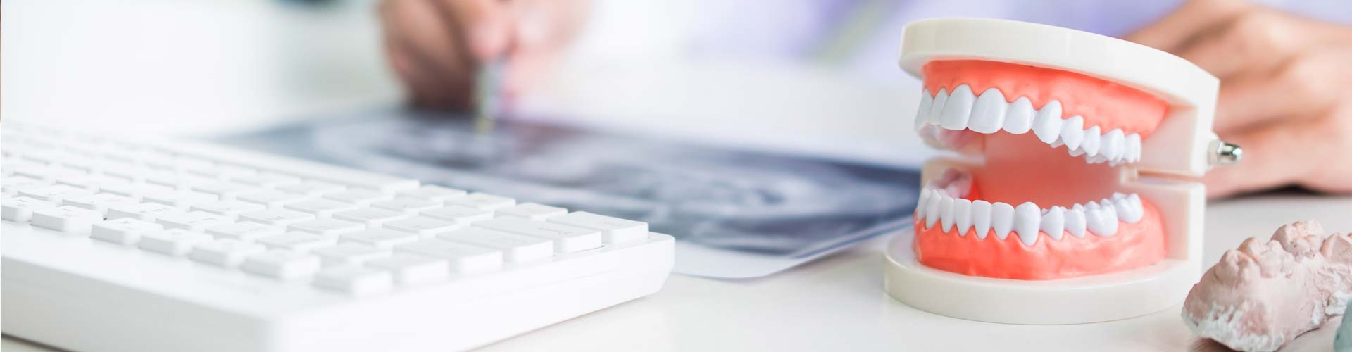
Apical periodontitis (AP) is an inflammatory disease of dental pulp origin, which will develop and continue to progress without obvious clinical symptoms. The choice of assessment methods for AP prevalence is controversial. Although histological analysis is still the gold standard for the diagnosis of AP, non-invasive imaging methods are more acceptable to patients in clinical practice. Therefore, radiographic examination to determine the existence of periapical light transmission zone is necessary for the diagnosis of AP.
Although there are many reports on AP, there is a lack of effective horizontal design to identify predictors of disease. Evaluating AP data at a separate level ignores that teeth exist in groups and are associated with patients. Each risk factor plays a different role. Therefore, it may increase the class I error rate and draw incorrect conclusions. At the same time, using this method to evaluate AP does not explain the variability of different factors, so it reduces the accuracy.
In order to explore the use of multifactor modeling in the study of AP related dental factors and patient factors, in October 2016, Hussein, from the school of Stomatology, National University of Malaysia, published an article in J endod to study the following problems: 1 Teeth have the potential for AP. 2. Is there any difference in the potential possibility of AP between patients. 3. When controlling other dental and patient factors, what is the relationship between each factor and the possibility of AP.
This study studied the random samples of several panoramic X-rays from the Stomatological Hospital of National University of Malaysia from 2011 to 2012. The periapical health status and root filling quality of imaging were evaluated by two examiners, and the periapical health status of each tooth was evaluated by Pai. Two examiners were calibrated with 10 digital curved slices that did not belong to the random samples of this study.
The two examiners observed the curved slice independently, and the consistency of the examiners themselves and between examiners was determined by Cohen kappa coefficient (k). The radiograph patients included in the analysis were 18 years old or older. If multiple radiographs of a patient can be used, only the earliest one should be used. In order to avoid overestimation of disease prevalence, the radiographs taken only for the diagnosis of AP were excluded. The radiographs of missing complete teeth were excluded. Use computer random numbers to generate random samples.
For multiple teeth, record the worst Pai score. Teeth with X-ray image in one or more root canals are classified as treated teeth. The root filling material terminates within 0 ~ 2 mm from the root tip on the radiographic image, and there is no visible gap, which belongs to just filling. In epidemiological studies, periapical index (PAI) is usually used to classify the degree of disease in imaging examination. Pai score greater than 2 is considered to be the manifestation of periapical disease. Exclude teeth that cannot be evaluated due to overlapping anatomical structures. In addition, if possible, apical radiographs were used to determine the existence of periapical transmission areas.
The study evaluated the curved slices of 233 patients, 147 women and 86 men. The age ranges from 16 to 70 years old. 93.1% of the patients had 20 or more teeth. AP was detected in 59 patients (25.2%), with 1 ~ 6 affected teeth per patient. A total of 6478 teeth, however, 69 teeth (without treated roots) were excluded due to image distortion or overlap.
The final 6409 teeth included in the analysis were 43 treated teeth. AP was found in 1.5% of untreated teeth (n = 96) and 37.2% of filled teeth (n = 16), with an overall prevalence of 1.7%. This study shows that the multifactor model considers the impact of many factors on disease, and is a comprehensive and effective statistical model for analyzing AP data.
Model 1 is a blank group, which believes that each tooth is independent, and the expected probability of AP is 0.42%; In model 2, dental factors are considered for prediction, and individual differences are considered. The differences between patients are statistically significant. The consistency coefficient within the group showed that nearly 53.16% of the variation was due to the differences between patients, and the remaining 46.84% of the difference was due to teeth or other unclear reasons; For the strict model (model 3), the posterior teeth are more likely to have AP than the anterior teeth, which may be due to the complexity of the anatomical structure. In addition, when examining periapical lesions, curved slices are less sensitive than apical slices, especially the anterior teeth, which may also be part of the reason.
In addition, the position of teeth, whether maxillary or mandibular, seems to have no effect. When other dental and patient factors are controlled, insufficient root filling greatly increases the probability of AP. In contrast, there was no significant difference between the filled teeth and the teeth without root canal treatment. There is a positive correlation between age and AP, which may be due to the accumulation of dental caries and multiple dental treatments. In addition, the relationship between gender and AP is not obvious.


 Open wechat
 | 粤ICP备2021067230号